 by: John Fedors
Hydrophilic spheres from Educational Innovations offer a variety of interesting applications and opportunities for scientific inquiry. They come in a variety of sizes: regular, jumbo, & gigantic. For the following examples, I prefer the regular or #710 size. However, whichever size you choose, they will expand to about 300 times their original dehydrated size.
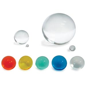 As they absorb the water, they become almost invisible, due to having the same refractive index as water. When placed in de-mineralized or distilled water and kept away from sunlight, they will dehydrate to their original size and can be re-used. Dehydration time will depend on air humidity.
Once enlarged, these clear spheres can be used to demonstrate:
* The lens of an eye (such as those of a shark, calf or sheep) that has the ability to magnify the print on a page. A thin slice may be used to mimic a cornea transplant.
* The suspension of small items such as a coin.
* Roots of a germinating seed.
Enlarged growing spheres can also help to observe the relationship of Surface Area (A=4pr2) to Volume (V=4/3pr3) mass in grams. They can be used to graph relationships.
Using dark vegetable dyes you can also relate to why living cells need to divide. The ratio of surface area to cell volume does not permit timely diffusion of required metabolites in or out of the cell. This can be demonstrated by placing a dyed sphere in clear water for 10 minutes and measuring the clear area of the sphere in relation to the rest of darkened or dyed sphere.
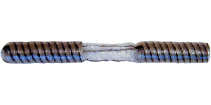
My favorite though is the demonstration of cell organelles/microstructures in eukaryotic cells. In addition to the hydrophilic spheres, this demonstration requires serpent skin tubing. Serpent skin tubing is a crinkled cellulose dialysis tubing that stretches out, remains open and relatively sturdy. It eliminates the usual wetting difficulty in opening traditional dialysis tubing.
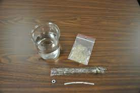
To demonstrate cell organelles/microstructures in eukaryotic cells with Growing Spheres you will need:
* Small nut & bolt (to serve as weight)
* Twist Ties (used in grocery produce departments or with some trash bags)
* Tall glass
* Food Dye
* Distilled or de-mineralized water
Here’s what you do…
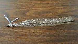
Take or cut a 6 to 8 inch length of Serpent Skin and flatten it. Fold it lengthwise 3 to 4 times, creating a long, narrow section. Fold the end up, then slide the folded end through the bolt. The bolt serves as a weight to keep the finished apparatus submerged in the dyed water.
Use a twist tie between the bolt and then end of the tubing. I am a fan of the champagne twist – twist six times as you would see the wire is twisted on a champagne bottle top.
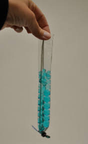
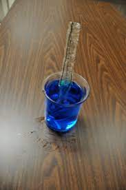 Place 25-35 Growing spheres through open end of the serpent skin and add 7-9 drops of dark vegetable dye to a tall glass. Add water to the glass up to an inch from the top or so. Place the weighted serpent skin with the growing spheres into the dyed water.
Results:
The dyed water will diffuse through serpent skin (cell membrane} and will cause the growing spheres to swell (this can take about 24 hours). The spheres will vary in size; larger spheres will collect towards the bottom of the glass while smaller spheres will collect towards the top. Adding more spheres initially will force them up and out. The varying sizes will help to visualize different organelles.
The dark stained organelles can be placed in clear colorless water for 5-10 minutes to demonstrate a colorless, clear outer surface area of diffusion. The spheres center will stay dark even after several water changes.
This also demonstrates the relationship of surface area to organelles volume and the need for the organelles to remain small for efficiency of passive diffusion.
